


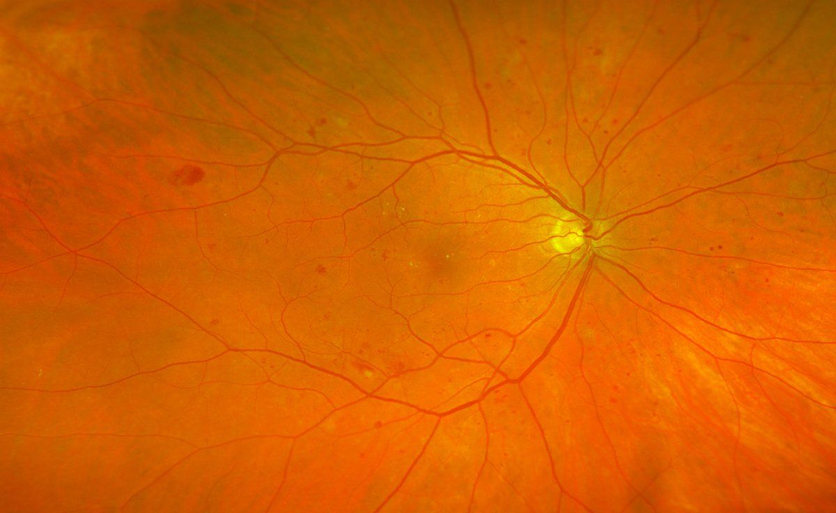
Findings from daily practice with the Optomap


Back to overview
Continue to:
Fluorescein angiographic images - optomap
tomographic images of the central retina - OCT
Laser-assisted tomography of an optic nerve head - HRT
Scheimpflug image of the anterior segment of the eye - Pentacam